Project Overview
We study physiological adaptations to natural and anthropogenic stressors in marine vertebrates and the role of oxidant stress and redox signaling in pre-clinical models of disease. Our overarching goal is to understand the physiological mechanisms that drive extreme adaptation in marine vertebrates and the contributions of redox biology in shaping the progression of several diseases. Current projects in our lab include 1) Stress and metabolic physiology in marine vertebrates, 2) Molecular mechanisms underlying diving-induced hypoxia tolerance, 3) the contributions of the antioxidant protein peroxiredoxin 6 to the maintenance of redox balance, and 4) the effects of dysregulated redox signaling in aging and metabolic disease.

Elephant seal dermal fibroblasts expressing GFP
Stress physiology
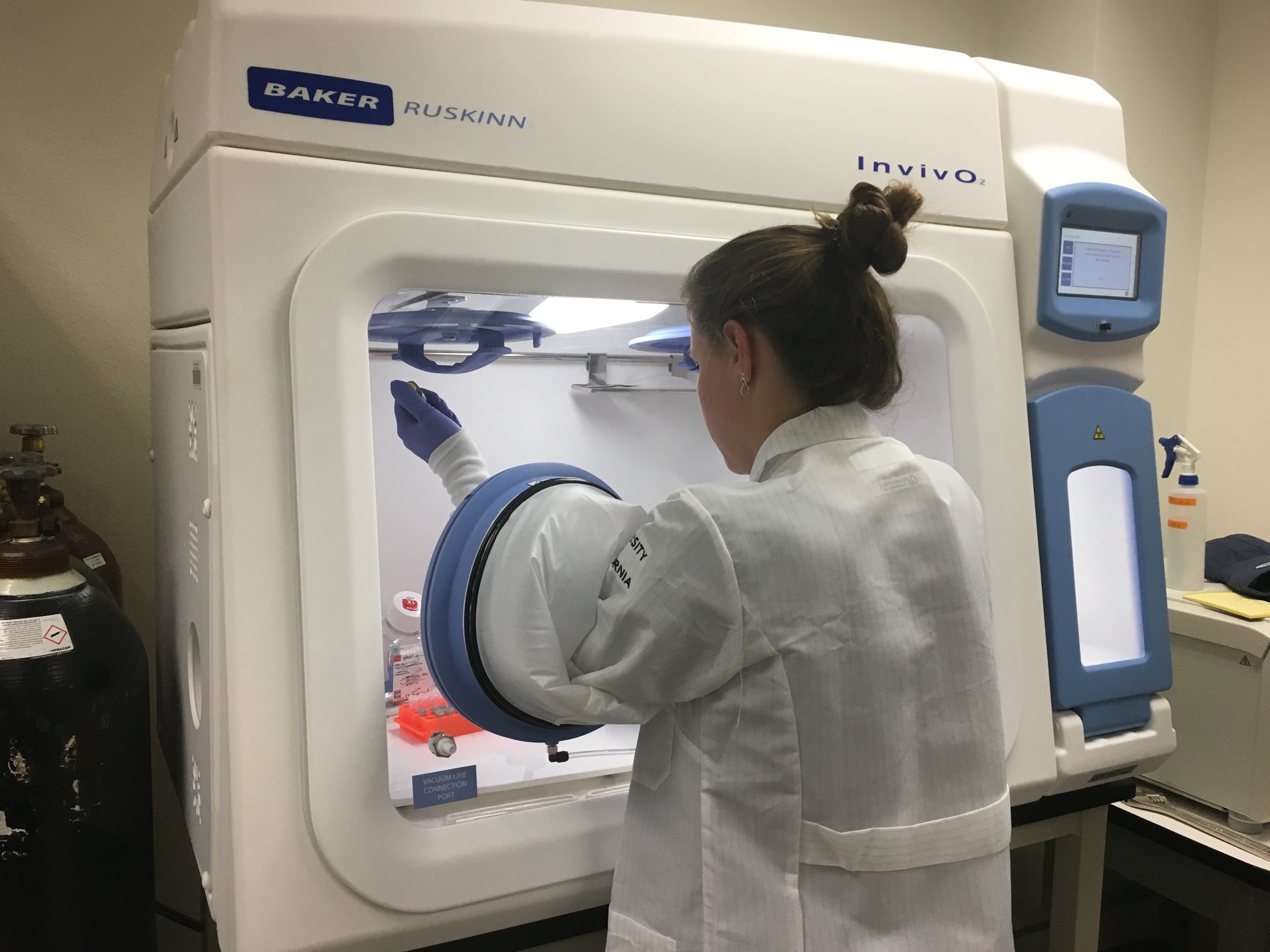
We are investigating the physiological consequences of exposure to natural and anthropogenic stressors in marine vertebrates using a combination of primary tissue culture models (myoblasts, fibroblasts, adipose-derived mesenchymal stem cells) derived from pinnipeds and cetaceans, and whole animal experiments with elephant seals and other marine vertebrates. These studies are a collaboration with several colleagues, including Dan Crocker (Sonoma State University), Tania Zenteno (CIBNOR, Mexico), Jorge Urban (UABCS, Mexico), Peter Sudmant (UC Berkeley), Jane Khudyakov (University of the Pacific), and Anders Goksøyr (University of Bergen). This work is supported by UC MEXUS, the Peder Sather Center, and the Norwegian Research Council, among others.
Selected publications:
Torres-Velarde JM, Kolora SRR, Khudyakov JI, Crocker DE, Sudmant PH, Vázquez-Medina JP. 2021. Elephant seal muscle cells adapt to sustained glucocorticoid exposure by shifting their metabolic phenotype. Am. J. Physiol. Regul. Integr. Comp. Physiol. 321: R413–R428. PMID: 34260302
Ensminger DC, Crocker DE, Lam EK, Allen KN, Vázquez-Medina JP. 2021 Repeated stimulation of the HPA axis alters white blood cell counts without increasing oxidative stress or inflammatory cytokines in fasting elephant seal pups. J. Exp. Biol. 224, jeb243198. PMID: 34524449
Lam EK, Allen KN, Torres-Velarde JM, Vázquez-Medina JP. 2020. Functional studies with primary cells provide a system for genome-to-phenome investigations in marine mammals. Integr. Comp. Biol. icaa065. PMID: 32516367

Catalytic sites of Prdx6
Peroxiredoxin 6
Peroxiredoxin 6 (Prdx6) is an antioxidant enzyme that expresses at least two different activities: glutathione peroxidase and phospholipase A2 in separate catalytic sites. We utilize knock-in mouse models with single-point mutations that inactivate each of the Prdx6 activities individually to investigate the contributions of this enzyme to the prevention of oxidative damage and cell death, as well as the maintenance of mitochondrial function. This work is supported by the National Institute of General Medical Sciences (Award Number R35GM146951).
Selected publications:
Arevalo JA, Xing D, Garcia-Leija R, Thorwald MA, Moreno-Santillán DD, Allen KN, Selleghin-Veiga G, Avalos H, Utke E, Conner JL, Brooks GA, Vázquez-Medina JP. 2025. Age-related declines in mitochondrial Prdx6 contribute to dysregulated muscle bioenergetics. Redox Biol. PMID: 40774144.
Torres-Velarde JM, Allen KN, Salvador-Pascual A, Leija RG, Loung D, Moreno-Santillán DD, Ensminger DC, Vázquez-Medina JP. 2024. Peroxiredoxin 6 suppresses ferroptosis in lung endothelial cells. Free Radic. Biol. Med. 218, 82-93. PMID: 38579937
Arevalo JA, Vázquez-Medina JP. 2018. The role of peroxiredoxin 6 in cell signaling. Antioxidants 7, 172. PMID: 30477202
Physiological hypoxia station
Hypoxia tolerance
Diving vertebrates are routinely exposed to hypoxia/reoxygenation due to the cardiovascular adjustments associated with diving. Since this is a natural behavior, they don't suffer from the classic complications observed after those events in humans (stroke, heart attacks, embolism). We are investigating the cellular and molecular mechanisms that underpin hypoxic and oxidant stress in diving vertebrates, utilizing primary cell systems cultured under controlled oxygen conditions, as well as in vivo experiments. Collaborators on this project include Allyson Hindle (UNLV), Celine Godard (Texas Tech), Peter Sudmant (UC Berkeley), and James Olzmann (UC Berkeley). This work is supported by the National Science Foundation, Office of Polar Programs (Award Number 2020664), the Winkler Family Foundation, and the National Institute on Aging (Award Number R21AG084993).
Selected publications:
Allen KN, Torres-Velarde JM, Vazquez JM, Moreno-Santillán DD, Sudmant PH, Vázquez-Medina JP. 2024. Hypoxia blunts angiogenic signaling and upregulates the antioxidant system in elephant seal endothelial cells. BMC Biology 23:91. PMID: 38654271
Arango BG, Ensminger DC, Xing D, Godard-Codding CA, Vázquez-Medina JP. 2025. Hypoxia exposure fine-tunes mitochondrial function in sea turtle cells. J. Physiol. 24, PMDI: 40991933.

White adipose tissue sections from lean and obese (OLETF) rats stained with perilipin.
Redox signaling in aging and metabolic disease
In collaboration with George Brooks (UC Berkeley) and Rudy Ortiz (UC Merced) we are studying the role of redox signaling and mitochondrial dysfunction in aging and metabolic disease. We use a combination of primary cell systems, aged mice, and rodent models of metabolic syndrome syndrome to conduct our studies. This work is supported in part by the National Institute on Aging (Award Number R01AG059715).
Selected publications:
Leija RG, Arevalo JA, Xing D, Vázquez-Medina JP, Brooks GA. 2025. The Mitochondrial Lactate Oxidation Complex: endpoint for carbohydrate carbon disposal. Am. J. Physiol. Endocrinol. Metab. 328, E126-E136. PMID: 39714986
Mendez DA, Hernández García J, Soñanez-Organis JG, Hernández Garcia M, Vazquez-Anaya G, Nishiyama A, Vázquez-Medina JP, Ortiz R. 2025. Exogenous Thyroxine Increases Cardiac Nrf2-TRX in Insulin-Resistant OLETF Rats. J. Endocrinol. 266, e250164. PMID: 40792604
Rodriguez R, Lee A, Godoy-Lugo J, Martinez B, Ohsaki H, Nakano D, Parkes D, Nishiyama A, Vázquez-Medina JP, Ortiz RM. 2021. Chronic AT1 blockade improves hyperglycemia by decreasing adipocyte inflammation and hepatic PCK1 and G6PC1 expression in obese rats. Am. J. Physiol. Endocrinol. Metab. 321: E714-E727. PMID: 34658252